Nagpur's Meditrina hospital docs do tracheal reconstruction, probably first in India

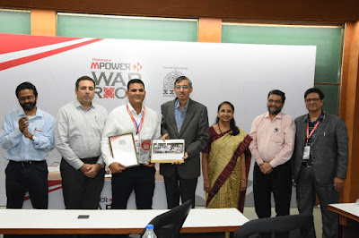
Dr Sameer Paltewar, Dr Parikshit Janai, Dr Gaurav Agrawal with the patient.
By Vikas Vaidya
IN A record of sorts, a team of doctors in city’s Meditrina Hospital has successfuly restored speech of a patient by implanting trachea reconstructed from the body part of the patient himself.
This is probably the first of its kind surgery performed in India.
The surgery was performed in four parts. The most difficult job was to implant the reconstructed trachea. It took 13 long hours but in the end the patient went home fit, fine and happy.
Girish, a city resident, had met with a road accident in May 2015. He was in a critical condition after sustaining polytrauma. He required long term intubation, tracheostomy and ventilatory support. After recovering from the injuries he developed tracheal stenosis, which is a known complication of long term intubation. As a result, he was not able to respire, not able to talk.
Girist visited a number of centers in India seeking treatment but returned disappointed. Two of the biggest problems he faced were inability to speak and constant possibility of blocking the tracheostomy tube.
Finally, Girish came to Meditrina Institute of Medical Sciences where he met Dr. Sameer Paltewar, a well-known Neurosurgeon.
Dr Paltewar discussed the case with Dr Parikshit Janai, Consultant Plastic Surgery, Microvascular and Cosmetic Surgery and Dr Gaurav Agarwal, Consultant ENT from Meditrina. It was decided to reconstruct his trachea by performing staged surgeries.
“Such extensive reconstructive surgeries of trachea are one of its kind and probably one of the first in India,” Dr Paltewar told ‘The Hitavada’.
First two stages involved opening the neck and knowing the size of wind pipe needed to be reconstructed. The 4.5 cm gap in the wind pipe was bridged with a temporary silicon T tube so that the patient could speak.
“It was successful but it was still a temporary solution. Accordingly a radial artery forearm flap was prepared by adding cartilages from both the ears (Prefabricated Radial artery flap). It was implanted in forearm to make it alive. After four weeks it matured. The cartilages were added to bring some rigidity to the tube made from his forearm skin,” he added.
In the third stage, two months after the first two, Girish’s neck was reopened. The surgery was difficult as doctors had to search proper blood vessels. His neck had been operated a number of times at other hospitals so it was in bad shape. Blood vessels were identified in the neck to receive the flap from the forearm. Skin from the forearm was harvested with the blood supplying vessels and a tube of 4.5 cm was created out of it.
“The tube was used to fill the gap between tracheal ends. The blood vessels of this ‘skin island’ were joined to the blood vessels in the neck to make this skin survive. This procedure was of critical nature and demanded exceptional clinical acumen coupled with unmatched operation theatre infrastructure. It took more than 12 hrs to complete this procedure,” explained Dr Janai and Dr Agarwal.
“Once we were sure that the tube we created is surviving and stable, a silicon stent was placed by Rigid Endoscopy and intervention. This made the tube more rigid and stable for the obstruction free breathing,” Dr Janai and Dr Agarwal told about the fourth stage.
Key elements in the entire procedure were the tertiary level readiness of OT, clinical acumen, and team effort.
According to Dr Janai, the contribution of team of anaesthesiologists was very important.
Team anaesthesia led by Chief Anaesthetist Dr Deepak Madankar, Dr Sheetal Samel, Dr Neelesh Mathankar, Dr Ritesh Borkar and Dr Latika Panpaliya had a unique challenge in managing this case as the wind pipe, which is the gateway for anaesthesia, was itself being repaired.
The team effort and one of its kind surgery bore fruits and Girish got his speech back. He responded well to the treatment and has since been discharged.
What is trachea
The trachea, commonly known as the windpipe, is a tube about 4 inches long and less than an inch in diameter in most people. The trachea begins just under the larynx (voice box) and runs down behind the breastbone (sternum).The trachea then divides into two smaller tubes called bronchi: one bronchus for each lung. Tracheal stenosis: Inflammation in the trachea can lead to scarring and narrowing of the windpipe. Surgery or endoscopy may be needed to correct the narrowing (stenosis), if severe.

Thanks Vikas Vaidya for sharing the beautiful Blog. Keep sharing more and more.
ReplyDeleteGreat site ........... For more details Visit Scholarships for Indian students studying abroad in the USA in Nagpur.
ReplyDeleteGreat Site. United Studies Abroad Consultants is offers mbbs in uzbekistan for Indian students.
ReplyDeleteGreat Site, USAC is the best for Admission consultants for study abroad programs in Nagpur. USAC is the only consultant that guarantees admission to MBBS, Bachelor's, Master's, MBA, or Ph.D. programs based purely on merit, without any donation.
ReplyDeleteGreat Site, Get the Admission consultants for foreign universities in Nagpur. USAC offers up-to-date guidance for students aspiring to study abroad, backed by years of experience in supporting individuals from diverse cultural backgrounds.
ReplyDeleteNice Information, Explore Bachelor's admission purely on merit in Nagpur. USAC is the only consultancy that ensures admission to MBBS, Bachelor's, Master's, MBA, or Ph.D. programs solely on the basis of merit, without any donation requirements.
ReplyDeleteNice Information, Explore Educations service consultant for US aspirants in Nagpur. USAC is the only consultancy that ensures admission to MBBS, Bachelor's, Master's, MBA, or Ph.D. programs solely on the basis of merit, without any donation requirements.
ReplyDeleteImpressive website! Looking for USAC, headquartered in Nagpur with over 14 branches in cities including Mumbai and Akola, has successfully placed more than 5,000 students in top universities worldwide.
ReplyDeleteFantastic website! Seeking for Admission consultants for foreign universities in Nagpur . Explore USAC Headquartered in Nagpur, operating over 14 branches across cities like Mumbai and Akola, USAC has successfully placed more than 5,000 students in prestigious universities around the globe.
ReplyDeleteThank you for sharing this informative post! It provided exactly the details I was searching for. A skilled anaesthesiologist plays a crucial role in ensuring safe and painless medical procedures. This post offers valuable insights to help patients make informed choices. Keep up the great work in highlighting the Anaesthesia Doctor in Etawah!
ReplyDeleteTop-notch website! Looking for Merit-based scholarships for Ph.D. admission in Nagpur. USAC's trained staff is committed to guiding students in pursuing their education abroad across top destinations, including Russia, China, the UK, the USA, Canada, Australia, and more.
ReplyDeleteGreat Blog. Get the best study abroad consultants nagpur. Visit USAC (United Studies Abroad Consultants) is a trusted education consultancy that helps students secure admission to top global universities based purely on merit, with no donation required.
ReplyDeleteGreat Blog. Looking for College admission counseling services in nagpur.USAC provides trusted overseas education services, ensuring guaranteed merit-based admissions for MBBS, Bachelor’s, Master’s, MBA, and Ph.D. programs without any donation.
ReplyDeleteFantastic website! Admission counseling for MBBS programs in Nagpur. For medical aspirants in Nagpur, USA Overseas is a trusted name. They provide specialized guidance for MBBS programs in countries like Bangladesh, Kyrgyzstan, and Russia, ensuring admissions to accredited universities with excellent reputations and affordable fees.
ReplyDeleteGreat to read about the achievements of Nagpur’s Meditrina Hospital doctors. For medical graduates inspired by such success stories, planning postgraduate studies abroad can be a smart move. Many are now opting for medical pg in germany, where tuition-free residency programs and structured training open doors to global career opportunities.
ReplyDelete